The Down and Dirty of Pet Dental Disease
Many Pets Harbor Hidden Painful Periodontitis
Or, How an Italian Greyhound Ends up Needing 17 Teeth Extracted...
Periodontal Disease refers to problems with the bone, gums, tough little fantastic periodontal ligaments inside the sulcus (tooth socket), basically everything around the tooth itself.
Doc Truli fills us in,”A careful exam of each tooth and 6 points of the gingiva around each tooth, and dental radiographs (x-rays) reveal hidden periodontal disease. There is no other way to find out what is wrong with your pet’s teeth. This examination requires general anesthesia, every time, every pet.”
Hidden Disease Doc Truli Finds Daily With X-Rays:
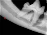
In the picture to your left, the root on the left sits in the white bone. The darker grey line in the center of the bone represents the marrow cavity. The bone is thinner, because marrow, nerves, and blood vessels live there. The root on the right is not resting in white bone, but rather, fuzzy, messy grey-black stuff. Pus and debris have eaten away the bone and the root sits in nothing good!
This x-ray was taken during a “routine dental” cleaning. The dog had no symptoms and the tooth looked fine from the outside. It did not wiggle in the jaw, because the left root held firm. Careful probing and an x-ray showed the massive problem.
If this dog had undergone non-anesthesia cleaning, this problem would persist. NO way would the person scaling the teeth have found this disease. The tooth was removed, and the dog gained a pound because she ate better after the surgery. This tooth was also a candidate for a tooth-sparing procedure in which the tooth could be cut in half, the good root could have a root canal, and a crown made to replace the diseased front half. In this case, the pet’s parent elected not to undergo the added expense and requested the disease simply be removed from the mouth.
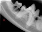
The little molar in the far left has an abscessed root and a large pocket. The technician scaled and polished the teeth after the full mouth radiographs were obtained. When she scaled the back molar, it was so loose and fragile, it fell out, leaving a hole in the jawbone. This tooth is categorized Grade 4/4 Periodontal Disease, and would’ve been extracted if it hadn’t fallen out on its own.
This hole was filled with a material to encourage new bone growth and the gingiva was delicately sutured in place to cover the sensitive hole. Antibiotics appropriate for the mouth and painkillers helped the dog through the healing process. Typically, Doc Truli recommends soft food for a time after the surgery (some veterinary dentists recommend 14 days of soft food).
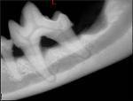
This dog had a loose abscessed back molar, too (Opposite side, so pocket is on your right.) Note how this molar sticks up out of the white bone. The black space under the top crown on the tooth in the furcation between the two roots should be filled with white bone just like in the tooth x-ray above. But it is not. This is called horizontal bone loss. It extends 25-30% down the root of the tooth. When it reaches 50%, that is Grade 4 out of 4 Periodontal disease, and the tooth will have to be extracted; it cannot be salvaged.
In this case, the tooth was a Grade 3 out of 4 Periodontal disease. Nothing will make the bone grow back in the furcation, but careful root planing to clean out any pus, calculus, and build-up can be performed, then the root can be treated to discourage further bone loss and disease. If the tooth was already loose, it would downgrade to a Grade 4/4, and surgical extraction would be the treatment option left to the pet.


great.. very informative!
I enjoy browsing your page, I usually learn random new facts.
Emily RandallHusky Training
Thanks Emily!