When a Fat Bump Goes Bad
Boxer Fatty Lump Goes Bad
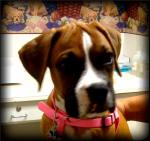
The first time I saw a fatty lump was also the day I saw my first convertible Jaguar XJ8 (before they sold to Ford.) The SUV in front of the pet emergency room, behind the Jaguar, held a nervous, frightened brown Brindle Boxer on a comforter, named Manny. Manny’s dad parked by the yellow curb and opened the overhead rear door to the SUV, placed the carpeted doggy ramp at the back car gate, and Manny slowly climbed down to the sidewalk. I saw the reason for the slow walking a few moments later. The late afternoon sunlight glowed across his Brindle side and then he turned.
The Boxer’s right side, where only his ribs should be, looked like a Basketball sticking out of his ribcage, covered with brindle fur.
Manny, the Boxer was diagnosed with a fatty lump — a Lipoma — several years earlier. Since the veterinarian said not to worry about it, the family did not worry as the thing grew and grew. Usually a lipoma shows up, almost as if instantly, is a certain size, and then stays that way. Sometimes a lipoma grows or shrinks if the dog gets fatter or skinnier. Generally, surgery is not needed.
In Manny’s case, because of the enormity of the lump, and the fact that Manny could not lie down to sleep on his right side, the lump was removed surgically for diagnosis and Manny’s comfort. It was a Liposarcoma.
Liposarcoma is a cancerous, metastatic, disease-causing tumor made up of fat cells.
Now, any lump that is just an -oma, is not so bad as a carcin-oma, or sarc-oma, both of which like to spread in the body and like to send microscopic satellites of themselves into the body, making them almost impossible to completely surgically remove.
The Boxer survived, but the massive surgery and the follow-up chemotherapy would have been unnecessary if intervention as soon as the lump started growing had been undertaken.
Sheltie Fatty Lump Goes Bad
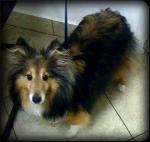
Nevada was the cutest, sweetest, most loved Shetland Sheepdog (Sheltie) I know. Nevada sported thick, shaggy fur in brown and white patches, with perfect white paws, brown soft fur on the top of his head, and his tongue hanging out in a perpetual smile. Usually, a visit to see the Doc was all licks and kisses.
“Doc, I’m worried about this lump I just found by his tail. It’s soft and it doesn’t hurt, but it keeps growing,” Nevada’s dad said over the phone.
“Bring Nevada right in,” Doc Truli said.
So there was Nevada four feet up in the air on top of an electric hydraulic lift table, butt facing the Doc, the lump looming about 4 inches in diameter next to the right side of his tail. Even with all of the fur, you could see the bulge.
Normally, an aspirate cytology will suffice for diagnosing a Lipoma (fatty lump). Aspirate cytology means sticking a needle into the lump, pulling back on the syringe, pulling the whole deal out of the lump, the pet parent saying, “Did you get anything?” and looking at the “nothing” in the barrel of the syringe. Then the Doc pfoofs the sample onto a microscope slide and the fat globs sparkle in the fluorescent indoor light. That’s usually all it takes.
Aspirate cytology means sticking a needle into the lump, pulling back on the syringe, and spreading the sample on a microscope slide.
The slide can be set aside to dry and be stained for analysis, but generally, the fat will not dry, the sample will not fix to the surface of the slide, and only a few fat cells, or nothing is left to look at under the microscope. Fabulous! A lipoma is diagnosed and everyone breaths easier.
There are a couple of glitches with this plan. One is, perhaps the sample came from a part of a tumor that is fat, but it is not representative of the whole lump; in that case, the diagnosis is missed altogether.
The other glitch is meanly serious. A Liposarcoma will reveal just fat under the microscope, too. It behaves in the body like a vicious cancer, but it is still made up physically speaking , of fat.
In Nevada’s case, yours Truli suspected foul play. The lump grew fast, was large, and everyone had a bad feeling about it. Instead of a plain old cytology, that would miss a sarcoma diagnosis, we performed a tru-cut biopsy.
A tru-cut biopsy involves a larger needle-like biopsy instrument that penetrates deeper and further than a skinny little syringe needle. It is designed to obtain a core sample. Lidocaine numbed the skin, a small incision allowed the tru-cut instrument to enter nicely under the skin, and then several core samples were obtained for histopathology analysis. The incision did not even need a stitch.
A tru-cut biopsy involves a larger needle-like biopsy instrument that penetrates deeper and further than a skinny little syringe needle. It is designed to obtain a core sample.
Nevada’s core sample looked like a squiggly worm piece of fat. The cores were dunked into formalin fixative and sent to the laboratory marked “stat.” We had a diagnosis of Liposarcoma within 48 hours. Nevada underwent surgery with a specialist oncologic surgeon- a surgeon that specializes in how to remove cancer without missing some or spreading it during the surgery.
Nevada felt good for about 10 months. Then he grew another lump on the left side of his tail and it grew fast. His parents decided not to make him undergo another surgery, and he felt okay with painkillers for another month. He larked in the yard and enjoyed spoiling with steak and treats. Nevada passed away 11 months after his original surgery. If he had been given only a cytology aspirate test and not a core tru-cut biopsy that first day, the Doc feels he would not have been around even a month, the tumor was growing so fast.
Bichon “Fatty Lump” Actually Masquerading Mast Cell Tumor
Snowy was a street dog. His mom found him hanging around her car, full of heartworms, matted, smelly, hungry, and friendly! She fed him and then brought him right in to see the Doc as soon as he finished his first meal.
“Doc, what kinda dog is he?”
Doc replied,”I think he’s a very dirty, mud-brown Bichon.”
“A what?,” his new mom asked.
“A Bichon. You know, usually a little white fluffy cute dog,” Doc said.
“That thing?” his mom fluffed up a little with pride,”Do you think he has papers?” **(You know– like registration, like AKC, or something…)
Education is one thing. Explaining the inexplicable is senseless. “Sure, he’s a real handsome guy under all this filth. We’ll clean up the fleas and ticks and dirt,” Doc said.
We explained the heartworm treatment protocol and cleaned up the little guy. He became known as Snowy after that first bath, not before!
Snowy was middle-aged, and he liked to eat. He grew a little fat and he grew lumps on his body that worried his mom. Like a good doggy mom, she had the lumps checked.
Now, a veterinarian can usually look at many different lumps and have a fair idea what they are, like adenomas. These are often hair follicle cysts, skin tags, or cauliflower-looking growths that are ugly, but won’t make your dog sick. The dreaded tumor is the mast cell tumor. It is made up of angry little immune cells called mast cells that release histamine when they are disturbed and can cause illness and even a shock reaction called anaphylaxis. And did I mention they come in three annoying grades of 1,2 and 3. Ones are removed surgically and you’re cured. Twos may or may not be cured, time will tell. And you had better have a thorough surgeon. Threes are nasty, have already spread to the internal organs, and surgery alone will not cure the problem.
The dreaded tumor is the mast cell tumor. It is made up of angry little immune cells called mast cells that release histamine when they are disturbed and can cause illness and even a shock reaction called anaphylaxis.
Mast cell tumors are the reason Doc Truli performs at least an aspirate cytology, measurement of the lump, and marks it on a dog body “map” in the medical record.
Snowy had 8 lumps on his body. 6 were adenoma-like skin bumps. Two were soft and under the skin. The one by his right shoulder aspirated fat only. The 1 cm lump in his right inguinal region (down between his legs on his lower belly), was full of mast cells.
Snowy went to surgery almost right away. Doc Truli removed the lump and 4 cm of normal-looking dog belly fat and skin all around the spot. About 11 stitches closed the whole thing, and a satellite dish and some painkillers helped Snowy at home the first day. After that, he did not care, and acted like he never had surgery.
Snowy’s lump was a Grade One mast cell tumor, cured by surgery.
Whew! That was a close one. If the Doc would have assumed that lump was fat, then it would have stayed a long time and has a chance to change and spread into a bad cancer.
P.S. If you were paying attention as you read, you may be asking yourself, “How does the doctor know if fat is from a sarcoma or a lipoma without the tru-cut biopsy test. Isn’t this just guessing?”
“Well yes,” says the Doc,”It is educated guessing. We know Liposarcomas are very rare and Lipomas are ubiquitous. Liposarcomas grow so fast the odds of seeing it when it is only 1 cm (half an inch or so) are tiny.”
Doctors have to make these kinds of educated guesses everyday.
P.S. (May 2010): Check out the “Bumps” tag or these posts to see other “bump” stories:
Beagle Bump Blues and Cocker Spaniel Belly Lump and Bichon Suffers Bump Under Tail


Trackbacks and Pingbacks