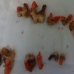
10-Year Old German Shepherd Dog Needs 9 Teeth Removed
This Shepherd’s Sewer Mouth Made Professional Nurses Faint!
Abigail’s mouth smelled like a toilet from across the examination room. The 10-year-old German Shepherd Dog looked normal. She acted normal. She even walked normally. But, if she licked your hand in greeting, you could not get that smell off your hand for 48 hours, no matter how many times you washed!
Abigail suffered horrible, horrible dental disease. This dog had one of the top ten worst mouths Doc Truli and the nurses had ever seen. Luckily, Abigail’s mom suspected there was a problem and came ready for dental surgery.
“I know her mouth is bad…” said mom.
(That’s worlds better than the usual shocked response Doc gets from pet parents, “How can it be that bad, he’s eating just fine.”)
Veterinary Technician Student Blown Away by Tartar
Doc Truli hosts clinical technician students from the local veterinary technician college. “Wow! My teacher would love to have those teeth so we can use them in class!” said the Thursday student.
“For what?” asked Doc.
“Our teacher mounts the teeth in a fake plastic dog jaw and we clean the teeth in the dental lab for practice,” said the student.
“Okay, well, I’ll ask Abigail’s mom if she wants to donate the teeth for teaching purposes after we get this mouth cleaned up,” promised Doc Truli.Veterinarians see many strange and disgusting things. Abigail’s mouth counts as one of those foul things we vow to fix.
Guided Tour of a Dog’s Tartar Removal
The tartar is so thick and crusted and caked onto the tooth surfaces that it looks like one big continuous shiny brown glop of muddy cement dentures. Underneath this filth and decay are delicate mucous membranes and tooth roots.

A horizontal ridge across the "middle" of the tooth structure demarcates the crown from the roots. Just as much root is showing as crown.
When the upper lip is lifted, you can see the tartar covering the upper carnassial tooth – the largest tooth in the dog mouth. The ridge across the tooth midway through the brown tartar is actually where the gingival margin should be. The gingiva, or gumline, has receded so much, the tartar covers the roots of the tooth, not just the crown enamel!
If you look at the previous picture, the calculus on the mandibular – bottom jaw – large back molar is also thick and extending too far into the gums. In fact, the x-ray of this molar reveals massive loss of the bone structure under the tooth.
X-Ray Showing Stage 4 Periodontal Disease in a Dog
The second of the 2 roots on the 2-root molar is sitting in a pocket of pus. On the x-ray, you see the crescent-shaped darker shaded area under the right side root? This is an area with no bone holding the tooth in place. Over 50% bone attachment loss is Grade 4 out of 4 Periodontal disease. This tooth had 100% attachment loss. Only the second molar root was holding the tooth in place. There is absolutely no saving a Stage 4 Tooth like this one; it must be removed from the body.
Abigail needed nine Stage 4 Periodontal Diseased Teeth extracted. Doc Truli removed the diseased, loose, infected teeth.
Tip for Strong Jawbone Healing: Consil
The tooth sockets were lavaged (cleaned with a clean water spray), and packed with Consil bone packing powder, pH balanced to be so alkaline no bacteria will grow in it, the Consil forms a matrix to speed new bone formation in the jaw.
“Research shows that sulci treated with the Consil powder regrow bone and the bone is just as thick as the normal bone at one-year radiographic follow-up examinations. What this means is, your dog’s jawbone will be stronger with the powder than without the powder,” says Doc Truli.
Abigail woke up uneventfully from her anesthesia. She asked for, and ate, a bowl of food 40 minutes after surgery! She continues to do well at home and her arthritis seems less painful now that Doc Truli removed the chronic dental irritation and inflammation.


